
Exploring Biominerals: Two (or Three) Photons Prove Superior to One
Suddenly, an intense pain starts in your back, under the ribs, radiating down to your abdomen. It’s probably a kidney stone (urinary lithiasis) which, once expelled, will require physico-chemical analysis. The aim is to determine the associated pathology, prescribe the right treatment and prevent recurrence. “Classically, urinary lithiasis is studied according to its morphology, or by infrared spectroscopy to determine its composition. However, this may not be sufficient to classify them effectively”, explains Marie-Claire Schanne-Klein, Research Director at LOB.
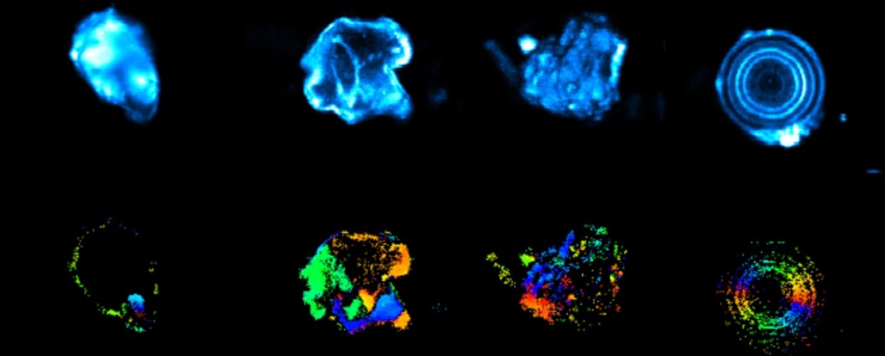
Multi-photon laser scanning microscopy, however, opens up a new avenue. A high-intensity pulsed laser probes deep into the kidney stones in a well-defined space (on a micrometric scale), providing valuable information on the arrangement of their molecules (crystalline structure), among other things. Thanks to a process of excitation by several photons reaching their target simultaneously, scientists observe either a fluorescence characteristic of the presence of proteins at a precise point in the sample, or signals specific to non-linear optics: second harmonic generation (SHG) under the action of two photons, or third harmonic generation (THG) with three photons**.
By combining its specialist polarimetry with SHG and THG, the LOB team is able to gain even greater insight into the internal structure of urinary lithiasis. Second harmonic results isolate certain crystalline kidney stones, i.e. those whose molecules are perfectly organized, with no center of symmetry. Third harmonic signals, on the other hand, generally reflect samples with interfaces between two media with different optical properties.
“We study kidney stone fragments, so there’s always a THG signal due to the air/lithiasis boundary. But polarimetry allows us to cross this barrier. A THG signal linked to birefringence can then be detected within the sample itself, typical of an anisotropic crystalline structure (editor’s note: whose physical properties vary according to the direction in which the object is viewed. In this case, the speed of light varies according to its direction of propagation) and therefore of certain classes of kidney stones”.
This innovative method is being extended to the analysis of various biominerals (kidney stones, calcifications, otoliths, etc.). Nephrologists are already interested in the results of this work, which could eventually lead to protocols and instruments that can be transposed to the hospital environment, closer to patients and doctors.