
New Insights into Tendon Regeneration: Lessons from Newts Hold Promise for Advancing Rapid Healing in Human Athletes
A research group led by Nagoya University’s Graduate School of Engineering has uncovered how rapid tendon regeneration occurs in newts. The research, published in the Journal of Orthopaedic Research, compared the regeneration mechanism of damaged tendons in newts with those in mice. In the future, their findings could help physicians to treat human athletes recovering from tendon injuries.
Tendon injuries are a serious obstacle for athletes, who currently require several months of treatment before they can return to competition. However, animals such as newts can rebuild severed tendons in as little as 12 weeks, without leaving scar tissue. To understand how they do this, a research group, led by Associate Professor Eijiro Maeda and Professor Takeo Matsumoto, investigated newts as a model for tendon regeneration. They chose the Iberian newt because it is one of the largest newts in the world, comparable in size to mice, making it easier to compare injury healing to that in mouse tendons.
“Mammals, such as humans and even mice, have a very limited healing capacity for organs and tissues,” said Maeda. “They repair damaged tissues with fibrous tissue (scar tissue) that differs from the original tissue. On the other hand, newts show an amazing capacity for regeneration of many types of tissues and organs without scar tissue formation. Therefore, findings from newt studies may show us how we mammals can heal/regenerate tissues and perhaps even organs.”
The researchers investigated injured flexor tendons from the middle toe of the hind foot, a body part common to both newts and mice. They found that six weeks after injury, the newts had developed new tissue resembling tendon tissue and at 12 weeks, the regenerated tissue showed strength comparable to that of a healthy tendon and was completely scar-free. However, in mice, the healing tissue differed from the healthy tendon tissue. Even at 12 weeks, its strength remained below that of the healthy tendon.
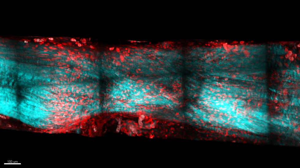
The researchers’ findings revealed that tendon-like collagenous tissue had bridged the tendon stumps in the newts. This differed from the disorganized scar-like tissue that filled the injured area in the mice. They also observed more cell nuclei around the junction of the newly formed and residual tendon in the newts. Since the number of cells are associated with cell growth, tracking them may be key to understanding the mechanism underlying regeneration of newt tendons.
“Although many studies have been conducted on tissue regeneration in newts and other amphibians, most of them have focused on large-scale regeneration such as missing fingers, limbs, and hearts,” Maeda said. “By focusing on relatively small-scale tissue damage regeneration, which can also occur in humans, we not only clarified the differences in healing and regeneration capabilities between newts and mammals, but also differences in the mechanisms of healing and regeneration.”
Maeda and his colleagues hope that their research will help reveal the mechanisms leading to full functional recovery of human tendon injuries. In the future, this knowledge may aid athletes, as well as other people suffering from tendon injuries, in recovering faster and returning to their previous lifestyle.
“The key to newt tendon regeneration is in the early stages after injury,” Maeda said. “They make a small and weak tendon first and it remodels, gaining strength and stiffness with time. If we can mimic this simple regeneration strategy, we can help human athletes to heal better without invasive surgery.”