UC San Diego Study: Liquid Droplets Influence Cellular Responses to Environmental Changes
Healthy cells respond appropriately to changes in their environment. They do this by sensing what’s happening outside and relaying a command to the precise biomolecule in the precise domain that can carry out the necessary response. When the message gets to the right domain at the right time, your body stays healthy. When it ends up at the wrong place at the wrong time, you can get diseases such as diabetes or cancer.
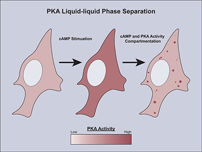
The routes that messages take inside a cell are called signaling pathways. Cells use only a few signaling pathways to respond simultaneously to hundreds of external signals, so those pathways need to be tightly regulated. New research by scientists at University of California San Diego has uncovered a surprising way that cells regulate signaling pathways. They found that when there are too many messages floating around inside a cell, the messengers form liquid droplets, sequestering themselves away where they can do no harm. The work was recently published in Molecular Cell.

“Liquid droplets organize cellular biochemical activities according to spatiotemporal regulation,” says Jin Zhang, Ph.D., professor of pharmacology at UC San Diego School of Medicine and senior author on the study.
The scientists worked with one of the main routes for cellular communication. It’s called the cAMP/PKA signaling pathway for its two main actors — cAMP (cyclic adenosine monophosphate) and PKA (cAMP-dependent protein kinase). When cAMP receives a signal from the cell’s surface, it activates PKA. PKA relays the message to the appropriate domain, whether it’s telling a specific gene to produce more protein or stimulating an enzyme to maintain a healthy level of glucose in the blood.
It’s not that simple, though. PKA carries messages to hundreds of different domains. According to Zhang, “At one moment, PKA needs to be active on the plasma membrane. But the next moment, it needs to come off the plasma membrane and be active on the mitochondrial membrane. Ten minutes later, it really needs to be in the nucleus to turn on transcription.”
To complicate matters, sometimes cells turn on too much cAMP and PKA. When that occurs, cell signaling becomes hyperactive and indiscriminate. Explains Zhang, “Different microdomains control different things. Let’s say you want the cAMP level to be high around calcium channels but low 10 nanometers away. How does the cell achieve that? By controlling cAMP.” But, she goes on, “PKA is the same. Typically, it’s recruited to specific domains by anchoring proteins. But if the PKA activity is too high, it will activate domains it’s not supposed to be activating. That’s loss of specificity.”
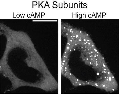
That’s why, according to the new research, cells form liquid droplets to ensure that the right message gets to the right domain at the right time. When the scientists analyzed the composition of the liquid droplets, as well as the timing of when they were formed, they found that cells formed the droplets using a subunit of PKA when too much cAMP and PKA were being turned on. In that way, the droplets sequestered the excess cAMP and PKA and tamped down non-specific signaling.
“We think that disappearance of the liquid droplets in Fibrolamellar Carcinoma (FLC) is one major contributor to hyperactive signaling that leads to tumorigenesis.”

In previous work, the authors found that a rare type of liver cancer called Fibrolamellar Carcinoma (FLC) blocks formation of these liquid droplets, resulting in uncontrolled cell signaling. “We think that disappearance of the liquid droplets is one major contributor to this hyperactive signaling that leads to tumorigenesis,” said Zhang.
FLC is a rare but devastating disease. It typically affects people under the age of 40 with healthy livers. The authors of this paper are hoping to investigate whether other cancers also cause a loss of liquid droplets and what the molecular mechanisms are behind it. Their ultimate goal is to design a molecular therapeutic to treat FLC — “anything,” says Zhang, “that helps us address the unmet needs of FLC patients.”
The authors of this study include Julia C. Hardy, Emily H. Pool, Jessica G.H. Bruystens, Xin Zhou, Qingrong Li, Daojia R. Zhou, Max Palay, Gerald Tan, Lisa Chen, Jaclyn L.C. Choi, Ha Neul Lee Dong Wang, Susan S. Taylor, Sohum Mehta, Jin Zhang at University of California San Diego and Stefan Strack at University of Iowa.
This work was supported by the National Institutes of Health and Fibrolamellar Cancer Foundation.

