
UCL’s Groundbreaking ‘Google Earth for the Human Heart’ Expected to Transform Cardiovascular Medicine
The study, published in Radiology, is an atlas of the human heart that captures the anatomical structure of the whole organ down to 20 micrometres – half the width of a human hair. In certain areas imaging has been done to cellular level.
The atlas will facilitate previously impossible research into both healthy and diseased hearts, clarifying anatomical structures and connections within the organ, with potential applications ranging from improving the treatment for arrythmia to creating more lifelike models for surgical training.
A showcase of the heart atlas and the technology behind it will be featured at The Wonders, part of the UCL Festival of Engineering, on Friday 19 July at 19:00 in the Bloomsbury Theatre.
Cardiovascular disease is the biggest cause of mortality worldwide. Ischaemic heart disease, a weakening of the heart caused by reduced blood flow, was alone responsible for 8.9 million or 16% of deaths globally in 2019, a figure that had risen by more than two million since 2000.
Clinicians typically use imaging techniques such as Ultrasound, Computed Tomography (CT) and Magnetic Resonance Imaging (MRI) to diagnose cardiovascular disease, but these techniques don’t provide detailed structural information about what’s happening in an organ. To get a more detailed view, it is necessary to slice organs into thin sections to be scanned, which significantly limits the field of view.
In recent years, a type of particle accelerator called a synchrotron has been used to develop new imaging techniques that overcome these limitations. Synchrotron studies on whole foetal and small animal hearts have been published, though these have always been at scales much smaller than major adult organs.
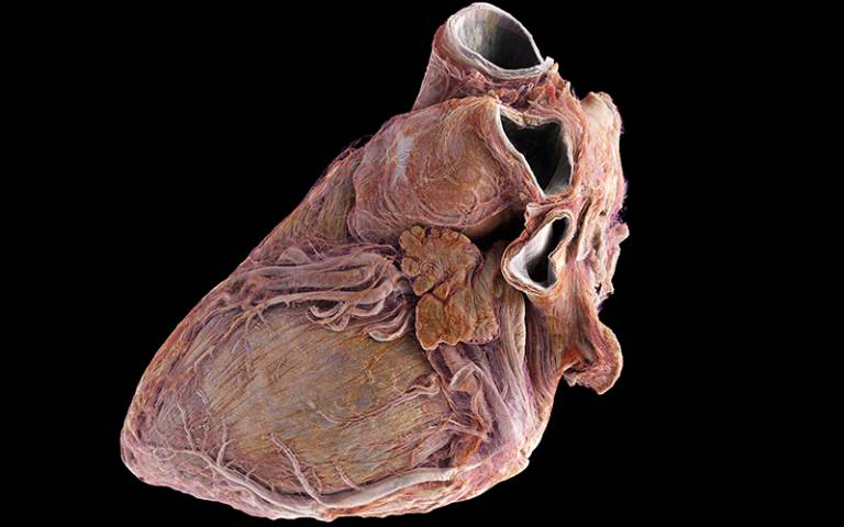
In this study, scientists from UCL and ESRF used an X-ray technique called Hierarchical Phase-Contrast Tomography (HiP-CT) to image two whole adult human hearts down to a scale of 20 micrometres, providing a comprehensive and detailed 3D view of the entire organ. One heart was from a 63-year-old White male donor without known cardiac disease (the control) and the other was from an 87-year-old White female donor with a history of ischaemic heart disease, hypertension, and atrial fibrillation. It wouldn’t be possible to image a living person’s heart in this way as the radiation dose would be too high.
Professor Peter Lee, senior author of the study from UCL Mechanical Engineering, said: “The atlas that we’ve created in this study is like having Google Earth for the human heart. It allows us to view the whole organ at global scale, then zoom in to street level to look at cardiovascular features in unprecedented detail.
“One of the major advantages of this technique is that it achieves a full 3D view of the organ that’s around 25 times better than a clinical CT scanner. In addition, it can zoom in to cellular level in selected areas, which is 250 times better, to achieve the same detail as we would through a microscope but without cutting the sample.
“Being able to image whole organs like this reveals details and connections that were previously unknown.”
The detailed imaging of the cardiac conduction system, which generates and transmits the electrical signals that drive the heart muscle’s pumping action, is one example of how the study will impact cardiovascular medicine.
Professor Andrew Cook, an author of the study and a heart anatomist from the UCL Institute of Cardiovascular Science, said: “With today’s technology, an accurate interpretation of the anatomy underlying conditions such as arrhythmia is very difficult. So, there is enormous potential to inspire new treatments using the imaging technique that we’ve demonstrated here.
“We believe that our findings will help researchers understand the onset of cardiac rhythm abnormalities and also the efficacy of ablation strategies to cure them. For example, we now have a way to determine differences in the thickness of tissue and fat layers located between the outer surface of the heart and the protective sac surrounding the heart, which could be relevant when treating arrhythmia.”
While the imaging of the two hearts is an important milestone for cardiovascular medicine, the researchers say that it will be necessary to image more hearts to get a more robust sense of the variation between individuals, taking into account differences in age, sex, ethnicity and disease progression.
The two hearts were imaged at the European Synchrotron Radiation Facility, which houses the world’s brightest X-ray source, situated in Grenoble, France.
Dr Joseph Brunet, first author of the study from UCL Mechanical Engineering and ESRF Visiting Scientist, said: “The first time you see the heart with HiP-CT it is quite surprising, as it clearly shows soft tissue not typically visible with conventional X-ray imaging. This is only possible because of the way that the phase contrast X-rays interact with these tissues, as well as the high energy that the ESRF can generate in order to penetrate the whole organ.”
This resolution is not without its challenges, though. Imaging for each heart generated 10 terabytes of data, one million times more than a standard CT scan.
Paul Tafforeau, an author of the study from ESRF who invented the HiP-CT technique, said: “The ESRF’s beamline facility is currently the only place in the world where complete adult human organs can be imaged with such a high level of contrast, and we are still quite far from the limits of the technology. The main limiting factor is the processing of the very large data produced by HiP-CT.”
This work contributes to the Human Organ Atlas project, which aims to establish an open science image database of all human organs in health and disease. To explore this data yourself visit human-organ-atlas.esrf.eu, or for more information on the Human Organ Atlas project see mecheng.ucl.ac.uk/HOAHub/ and videos at bit.ly/HiP-CT-Heart.
The project is co-funded by the Chan Zuckerberg Initiative (CZI).