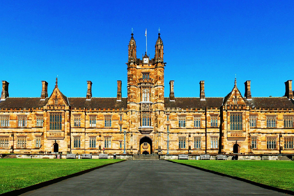
University of Sydney: Preserving a body of work
For over 135 years, the University of Sydney has held a special place for the preservation of pathology specimens. The collection is a fascinating and invaluable resource for students, educators, and researchers, and has even inspired artworks and literature.
Now, thanks to generous donors, this collection is being cared for and digitised in perpetuity for future generations, tucked away behind a discreet door off the foyer of the Charles Perkins Centre.
1888 flask of broth made and sealed by Louis Pasteur
Among the 1500-plus specimens are rare pieces including a heart dating back to 1895, a lung infected with the deadly ‘Spanish flu’, and medical instruments used since the Sydney Medical School began teaching in 1883. One of the most prized exhibits is an original 1888 flask of broth made and sealed by Louis Pasteur.
Essential for seeing firsthand the impacts of disease on human organs, this collection has long been used by students, including Dr Edward “Ted” Kremer OAM (MBBS ’70).
Having studied medicine in the 1950s, he has always been captivated by historical instruments and specimens. Wanting to assist in securing its future, he asked his network if they would support the collection through philanthropy. Dr Len Ainsworth AM was one of those people.
“As a former medical student, and following further discussion, I said to Ted it would be my pleasure to help provide enduring resources to enable the collection to be maintained long term. I have a keen interest in medical research in quite a number of areas and understand and agree with the importance of the pathology collection and its long-term activities,” Ainsworth said.
The collection is also maintained with the help of the Susan and Isaac Wakil Foundation, the Glendonbrook Foundation and the Hunt Endowment.
Musicians rehearse at the Sydney Conservatorium of Music
Dr Murat Kekic (MSc ’96, PhD ’04) holding a diseased heart specimen
Over the last twenty years, the collection has grown and expanded under the skillful hand of its curator, Dr Murat Kekic (MSc ’96, PhD ’04). Having majored in biotechnology, he worked in the University’s Museum of Human Anatomy and then went on to do his PhD in muscle proteins.
For Kekic, there is no average day. One day, he might collect tissue taken from autopsies or surgical procedures. The next, his team will preserve the specimens, constructing the perspex containers, fixing the items in place and adding preservation fluids.
With a mix of awe and inquiry, Kekic points to a hairball that’s taken on the shape of the stomach of a habitual hair eater; a teratoma (tumours that have developed teeth and hair); and a black lung, which is shockingly dark, taken out of a coal miner. As learning tools, they are irreplaceable. The black lung has helped inform work, health and safety processes, as it shows first-hand what happens without laws in place.
For medical students, the need to know what diseases look like and how organs function while afflicted is critical. If you haven’t seen it, you can’t recognise it. This is where the digitisation steps in, aided by Jim Cook, the Acting Manager of Digital Innovation Strategy.
Cook muses that his role is to “help our academic friends into the light of tech innovation.” An off-the-shelf solution couldn’t be found, and this gift has enabled the team to create a unique technological platform – Etaki.
Etaki is a digital content management system to manage virtual reality, 3D and gigapixel content as well as microscopic, macroscopic and tomographic representations, all in one centralised, secure and accessible platform. The team used laser active scanning, MRI, and CT technology to digitise objects.
The cloud-based solution provides an equitable experience for students who can’t visit the museum in person, and also means students no longer need to download enormous files. The next stage will expand into virtual reality and artificial intelligence.
Specimens on display
click to show image
click to show image
click to show image
click to show image
click to show image
click to show image
click to show image
next slide
previous slide
“This enhanced experience is as close as we can get to stepping inside a living body,” says Cook. “As an example, a heart specimen is only as big as a fist – it’s hard to see at close range. With a high-resolution digital image, you can zoom in very close.” Tutors can also annotate specimens, enabling a consistent and high-quality interactive experience for students.
“Ten years ago, teaching flipped to focus on real-world applications and interaction beyond the lecture theatre. Technology plays a vital role in preparing our students for their future work and creating graduates who are leaders in their field.”
Cook’s favourite feature is the ability to add sound, and he’s looking forward to students hearing the difference between healthy and unhealthy lungs breathing. Or hearing a sedentary heartbeat compared to the heartbeat of an elite athlete while exercising.
The 3D models show these organs in situ to garner a greater understanding of how they work together. Generally, students’ only experience with real organs is through cadavers, whose lungs are deflated. Digital models can show the full range of motion and the impact inflation and deflation has on other organs.
Opportunities for research are expanding too, with the technology offering newfound capability to share the collection with other universities while adhering to legislative requirements. Timely indeed: with renewed interest – encouraged by the coronavirus – to understand the pathologies of the past.
One of the rarest items – a lung infected with ‘Spanish flu’ – is drawing the attention of researchers from across the globe, as this specimen is the most complete known sample. Researchers can take a slice from the organ and extract genetic material to chart the history of the virus over many years.
“We can learn a lot from the past about how things evolve,” says Kekic. “Even over the last 100 years, there have been staggering advancements in medicine. This is a capsule of the past that’s informing the future.In 100 years from now, imagine what will be possible.”
It is striking to think what these vital learning aids will enable for future generations, thanks to the generosity of forward-thinking donors today.

